
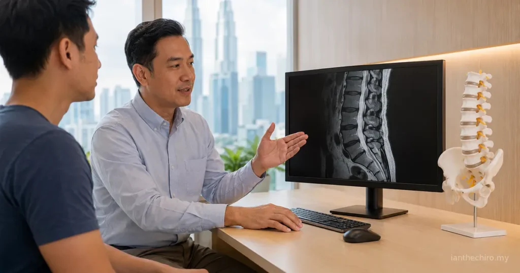
Your back has been painful for two weeks. It catches when you stand, feels stiff after driving and has not settled as quickly as you expected. A friend says you should get a scan before anyone touches it. So, do I need an MRI for back pain, or can an assessment come first?
For most people with a new episode of uncomplicated back pain, an MRI is not the first step. The decision depends on the symptom pattern, neurological findings, medical history and whether the result would change what happens next. Our back pain, slipped disc and sciatica guide explains why similar symptoms can come from different patterns.
At a Cheras clinic, patients often arrive worried that waiting for an MRI means ignoring the problem. It does not. A careful history and examination can identify whether conservative care is reasonable, whether imaging may be useful later, or whether medical assessment is needed promptly.
Quick Answer: Do I Need an MRI for Back Pain?
You usually do not need an MRI immediately for back pain that started recently, has no concerning warning signs and is not accompanied by progressive neurological loss. Many episodes can be assessed and managed initially through history, physical examination, appropriate activity and review of progress.
An MRI becomes more useful when there is concern about a serious underlying condition, when nerve function is worsening, when symptoms remain disabling despite appropriate care, or when a specialist is considering an injection or operation and needs anatomical detail.
The useful question is not simply whether an MRI can find something. It is whether the finding is likely to explain the current pattern and change the next decision.
What Can a Lumbar MRI Show?
MRI uses a strong magnetic field and radiofrequency energy to create detailed images. It does not use ionising radiation. A lumbar MRI can show structures that are not seen clearly on an ordinary X-ray, including:
- intervertebral discs and disc hydration
- disc bulges, protrusions or extrusions
- the spinal canal and spaces where nerves travel
- nerve-root compression or crowding
- ligaments, muscles and other soft tissues
- bone marrow changes
- some features of inflammation, infection or tumours
This detail can be valuable when the clinical question is specific. For example, if a person has persistent leg pain, numbness and objective weakness that fit one nerve-root pattern, an MRI may help a specialist confirm which level is involved before planning a procedure.
However, MRI is not a live video of pain. It cannot show how much a finding hurts, measure fear or sleep disruption, or prove that every visible change is responsible for the symptoms. A clear image is not automatically a clear diagnosis.
MRI reports also use descriptive language that can sound more alarming than intended. “Degeneration” describes structural ageing. “Bulge” describes the shape of a disc extending beyond its usual boundary. “Stenosis” means narrowing, but the report may grade it as mild, moderate or severe and identify whether it affects the central canal, a side recess or the nerve exit. None of these words should be interpreted without asking where the symptoms are and whether neurological findings support the same level.
The radiologist interprets the images and answers the clinical question provided on the referral. If that question is vague, the report may list many features without identifying which one matters. The quality of the referral question affects the usefulness of the report. This is another reason an assessment can be valuable before imaging.
Why MRI Findings and Back Pain Do Not Always Match
Disc degeneration, bulges, small protrusions and facet-joint changes become more common with age. Many people have one or more of these findings without back pain.
A widely cited systematic review of spinal imaging in people without symptoms found that degenerative features were common even among pain-free adults and increased with age. The practical lesson is not that scan findings never matter. It is that they must be interpreted alongside the symptom route, examination and clinical context.
Two opposite mistakes can follow an MRI:
- treating every visible change as dangerous damage
- dismissing a relevant finding merely because similar changes can occur without pain
The better approach is correlation. Does the side, level and type of finding fit what the person feels? Do strength, sensation or reflex changes support it? Would the result alter care?
Your spine is not a car part that fails because a scan uses the word “degeneration”. Many imaging terms describe age-related structural change rather than a deadline for surgery.
When an MRI Is Usually Not the First Step
Routine early MRI is usually not recommended for uncomplicated low back pain with or without mild leg symptoms when there are no red flags and no progressive neurological findings.
The American College of Radiology low back pain criteria rate lumbar imaging as generally inappropriate at the start of uncomplicated acute low back pain. This is because early imaging often does not improve the initial decision and may reveal incidental findings that create extra worry or unnecessary procedures.
Common situations where an immediate MRI may not add much include:
- pain began after ordinary lifting, sitting, training or an unfamiliar activity
- movement changes the pain, but there is no major trauma
- there is no progressive weakness, widespread numbness or loss of coordination
- bladder and bowel function are normal
- there are no strong signs of infection, cancer or fracture
- symptoms are already stable or gradually improving
This does not mean “nothing is wrong”. It means assessment and a monitored initial plan may provide more useful information than scanning everyone on day one.
When Might an MRI Be Appropriate?
MRI is more likely to be useful when the result could change referral, treatment planning or urgency. The decision should consider the full history rather than one symptom in isolation.
Serious underlying conditions are suspected
MRI may be requested when there is concern about infection, cancer, inflammatory disease, fracture complications or compression of the nerves at the bottom of the spinal canal. The exact imaging pathway may differ, and hospital teams may choose MRI, CT or another test depending on the suspected condition.
Neurological function is worsening
Progressive loss of strength, worsening foot control, expanding numbness or significant changes in reflexes can justify earlier medical investigation. A person who is simply hesitant because movement hurts is different from someone whose muscles repeatedly cannot produce expected force during testing.
Persistent nerve-root symptoms affect decisions
An MRI may be considered when leg pain, tingling, numbness or weakness continues despite an appropriate period of conservative care and the result would guide specialist review, an injection or surgical planning. The article on why sciatica can feel worse when sitting explains how a nerve-related pattern may behave before imaging is considered.
Persistence alone is not enough. A person whose leg pain has lasted eight weeks but is steadily improving, walking further and regaining sleep may not have the same imaging need as someone whose symptoms remain severe, function is worsening and examination shows a consistent nerve deficit. The direction of change matters alongside the calendar.
Similarly, an MRI may be reasonable when the diagnosis remains uncertain after a thorough assessment and the uncertainty is blocking an important decision. It should not be used merely to reassure someone that every structure is perfect, because age-related findings can create new worry even when no dangerous cause is present.
A procedure or operation is being considered
When a clinician is deciding whether an intervention is suitable, imaging can help identify the relevant level and anatomy. Even then, the scan must match the clinical findings. The presence of a disc bulge alone is not a procedure plan.
Which Back Pain Symptoms Need Urgent Medical Assessment?
Most back pain is not caused by an emergency, but some combinations should not wait for a routine clinic appointment.
⚠️ Seek urgent medical assessment if back pain occurs with new loss of bladder or bowel control, numbness around the groin or saddle area, rapidly worsening leg weakness, major loss of balance or walking ability, fever or severe illness, significant trauma, a known cancer history with new unexplained symptoms, or severe unrelenting pain that is rapidly worsening.
Hospital clinicians will decide which tests are needed. MRI may be important in some of these situations, but the priority is urgent assessment, not arranging a private scan and waiting for a report.
If you are unsure whether your symptoms need medical review first, the Ian The Chiro symptom checker can help you identify a safer starting pathway. It is not a substitute for emergency care.
Do Leg Pain, Numbness or Tingling Mean You Need an MRI?
Not automatically. Pain travelling into the buttock or leg can occur with nerve-root irritation, but it may also arise from muscles, joints, tendons, the hip or peripheral nerves. Tingling can strengthen the suspicion of nerve involvement without proving the cause or level.
Assessment looks for a pattern:
- whether symptoms travel below the knee
- which part of the leg or foot is affected
- whether coughing, sitting or spinal position changes the symptoms
- whether sensation differs between sides
- whether specific muscle groups are genuinely weak
- whether reflexes have changed
- whether the pattern is stable, improving or worsening
The guide to numbness in the leg and lower-back-related symptoms explains why the route and neurological findings matter. When these findings are severe, progressive or persistent enough to change the next step, MRI becomes more relevant.
How Long Should You Wait Before Getting an MRI?
There is no universal countdown. “Six weeks” is often mentioned because many uncomplicated episodes improve during the first several weeks and imaging is unlikely to change early care. It is a guideline, not a rule that everyone must wait.
Earlier imaging may be appropriate when red flags or meaningful neurological loss are present. Later imaging may be reasonable when symptoms remain disabling despite a well-matched plan, the diagnosis is uncertain, or a specialist needs the result for an intervention.
What matters during the waiting period is not passive delay. There should be a clear plan for activity, symptom management and review. A review point is more useful than an arbitrary scan date. If strength, sensation, bladder or bowel function changes, the plan should change sooner.
MRI vs X-Ray vs CT: What Is the Difference?
The best test depends on the question.
MRI
MRI is particularly useful for discs, nerves, the spinal canal, bone marrow and other soft tissues. It takes longer than an X-ray or CT and requires the person to remain still. Some people find the enclosed space difficult. Metal implants, implanted devices, pregnancy and kidney function may affect how the scan is planned, especially if contrast is being considered.
X-ray
X-rays show bone alignment, fractures and some degenerative changes but do not show discs or nerves in the same detail. They use ionising radiation. Routine X-rays are not required before every chiropractic visit, as explained in whether you need an X-ray before seeing a chiropractor.
CT
CT uses X-rays to create detailed cross-sectional images. It can be particularly helpful for bone and trauma assessment and may be used when MRI is unsuitable or unavailable. It exposes the person to more ionising radiation than a standard X-ray.
More advanced does not mean more appropriate. A test is useful when it answers the clinical question with a reasonable balance of benefit, burden and risk.
Ultrasound has useful roles for some superficial soft tissues and abdominal or pelvic questions, but it cannot provide the same view of lumbar discs, the spinal canal and nerve roots as MRI. Bone scans and blood tests may occasionally be used when infection, inflammation or abnormal bone activity is suspected. These are not interchangeable “better scans”. Each belongs to a different diagnostic pathway.
In Malaysia, availability and cost can influence where imaging is performed, but convenience should not replace indication. Before paying for a scan, ask who will interpret it, whether the correct spinal region and protocol have been requested, and what decision will be made differently once the result is available.
What Should Be Assessed Before Imaging Is Ordered?

A good assessment does not attempt to “beat” an MRI. It answers a different set of questions that imaging cannot answer alone.
History and symptom behaviour
The clinician should ask how the pain began, where it travels, which positions alter it, whether sleep or function has changed and whether any medical warning signs are present. Past cancer, infection risk, trauma, osteoporosis, medications and previous spinal procedures may matter.
Movement and loading
Different movements, positions and tasks can show whether symptoms are mechanically responsive. The aim is not to force pain or prove one damaged structure. It is to identify safe movement, meaningful restrictions and patterns that can be monitored.
Neurological screening
Strength, sensation, reflexes, walking and coordination may be checked when nerve involvement is possible. Findings should be interpreted together. One uncomfortable strength test is not the same as a consistent neurological deficit.
Baseline and review criteria
A useful initial plan records what the person can currently do and what would trigger reassessment. That may include walking tolerance, sitting time, symptom distribution, sleep, strength or work capacity.
What If the MRI Is Normal but Your Back Still Hurts?
A normal or reassuring MRI does not mean the pain is imaginary. MRI is designed to show anatomy, not every contributor to pain.
Pain can be influenced by tissue sensitivity, movement tolerance, muscle capacity, sleep, stress, previous episodes and how the nervous system responds to threat. Some painful structures or functional patterns may not produce a dramatic scan abnormality.
The next step is to reconsider the clinical pattern rather than chase progressively more tests without a clear question. A clinician may review the hip, sacroiliac region, peripheral nerves, load tolerance or non-musculoskeletal causes depending on the presentation.
Pain deserves assessment even when the scan does not provide a neat label. The goal remains improved function and a safe plan, not forcing the report to explain every sensation.
What If the MRI Shows a Disc Bulge or Degeneration?
First, do not read the report as a verdict. Terms such as disc bulge, desiccation, degeneration, annular fissure and facet arthropathy describe visible features. Their importance depends on whether they match the symptoms and examination.
A disc finding is more clinically persuasive when the side and level fit a consistent nerve-root pattern. It is less persuasive when the report lists several levels but the symptoms do not match any of them.
Many disc-related episodes can improve without surgery. If an operation is being discussed, the guide does a slipped disc need surgery explains why symptoms, neurological function, response to care and personal goals matter alongside imaging.
Do not repeatedly test painful movements because the report sounds severe. Equally, do not ignore worsening weakness because someone said disc findings are always normal ageing. Context changes the meaning.
Does Having an MRI Change Back Pain Treatment?
Sometimes, but not always.
An MRI may change management when it identifies a condition needing medical or surgical care, clarifies a persistent nerve-root problem before a procedure, or rules in or out an important differential diagnosis.
For uncomplicated back pain, the initial conservative plan may remain similar whether or not a scan shows age-related changes. Education, gradual activity, exercise matched to capacity and symptom review can still matter. Manual treatment may be considered only when appropriate and should support a broader active plan rather than become automatic.
An MRI can still have value even when it does not lead to surgery. It may help a specialist exclude a suspected condition, refine a diagnosis or discuss realistic options. Conversely, a striking report may not change care if the person is improving and the finding does not match the examination.
Useful questions after an MRI include:
- Which finding, if any, matches my symptoms?
- Are there neurological findings that support the same level?
- Is anything urgent or medically important?
- What options would be different because of this result?
- What should be monitored if conservative care continues?
A scan should support shared decision-making, not replace it. If no one can explain how the result changes the plan, the practical value of the scan may be limited.
Dry needling targets selected muscle-related contributors. It does not treat a disc bulge shown on MRI. Adjustments or mobilisation aim to influence comfort and movement; they do not push a disc back into place. Exercise develops tolerance, strength and confidence over time. These options should remain distinct.
What Can You Do While Monitoring Back Pain?
If urgent features have been excluded and an initial conservative approach is reasonable, the goal is usually to keep life moving within tolerable limits.
- change position regularly rather than searching for one perfect posture
- continue comfortable walking and ordinary daily activity
- reduce a clearly provocative load temporarily instead of stopping everything
- avoid repeatedly bending or twisting just to “check” whether it still hurts
- track whether pain is becoming more local, less intense or less limiting
- note any new numbness, weakness, coordination or bladder and bowel change
- follow medication advice from a qualified medical professional
Long periods of bed rest can reduce confidence and capacity. A flare after activity does not automatically mean damage has worsened, but a progressively changing neurological pattern should be reassessed.
How Ian The Chiro Approaches Back Pain and Imaging
At Ian The Chiro, the first step is assessment, not ordering a scan by default or promising that treatment will solve every scan finding.
The consultation considers the symptom history, movement, relevant neurological findings, daily demands and warning signs. Existing MRI, X-ray or CT reports can be reviewed in context. If the presentation appears suitable for conservative care, the plan may combine education, activity guidance, exercise and selected hands-on care.
If the findings suggest that imaging or medical review could change the next step, referral is more appropriate than continuing routine treatment. The aim is to use MRI when it answers a meaningful question, not as a ritual before care begins.
When Should You Book a Consultation?
Consider an assessment when back pain persists, keeps returning, limits normal activity or includes leg symptoms that are difficult to interpret. You do not need to arrange an MRI before booking unless a medical professional has already advised it.
If you are in Cheras or Kuala Lumpur, a chiropractic consultation can help determine whether conservative care appears suitable, whether progress should be monitored first, or whether medical referral and imaging make more sense. Urgent warning signs should go directly to medical care.
Do I Need an MRI for Back Pain? Frequently Asked Questions
Can an MRI tell exactly what is causing my back pain?
Not always. MRI can show discs, nerves and other anatomy, but common findings may occur without symptoms. The report must be matched with the pain pattern, neurological examination and clinical history.
Should I get an MRI before seeing a chiropractor?
Usually not for uncomplicated back pain. A consultation can identify whether imaging is likely to change the next step. Bring any existing scan and report, but do not arrange a new MRI solely because you assume it is required.
Is MRI safer than an X-ray?
MRI does not use ionising radiation, while X-ray and CT do. That does not make MRI automatically better for every problem. Each test answers different questions and has its own limitations and suitability considerations.
Does sciatica always need an MRI?
No. Mild or improving sciatica can often be assessed and managed initially without imaging. MRI becomes more relevant when neurological loss is progressive, symptoms remain disabling or a specialist needs anatomical detail for an intervention.
Will an MRI show a trapped nerve?
It may show narrowing, disc material or another structure contacting a nerve root. The finding is most meaningful when it matches the symptom distribution and neurological signs. Contact on a scan does not always mean the nerve is causing pain.
What if I am claustrophobic?
Tell the referring clinician and imaging centre. They may discuss preparation, a wider-bore scanner or other options. Medication for anxiety must be prescribed and may affect driving arrangements.
Do I need contrast for a back MRI?
Most routine lumbar MRI examinations for back pain do not require contrast. Contrast may be considered for specific questions such as infection, tumour or some postoperative problems. The referring and radiology teams decide based on the clinical question.
If you have kidney disease, an implanted device, metal fragments, previous spinal surgery or a possible pregnancy, tell the referring clinician and imaging centre before the appointment. The radiology team will confirm whether the planned scan and any contrast agent are appropriate.
Can I exercise before getting an MRI?
If serious pathology has not been suspected, comfortable activity and appropriately scaled exercise are often reasonable while symptoms are monitored. Stop and seek reassessment if exercise triggers spreading neurological symptoms, worsening weakness or another concerning change. The guide on whether to rest or keep moving when something hurts offers a practical framework.
When should persistent back pain be reassessed?
Reassessment is sensible when symptoms are not improving as expected, continue limiting work or sleep, spread into the leg, or occur with numbness or weakness. New urgent warning signs require immediate medical assessment rather than a routine review.